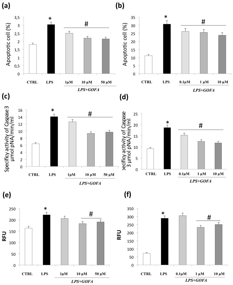
Figure 5.
GOFA effects on apoptosis and cell senescence. The effect of GOFA on apoptosis of U937 (a) and HCT116 (b) cells. The cells were treated with indicated concentration of GOFA for 1 h and then exposed to LPS for 24 h. An Annexin V assay was used for apoptosis detection. (c) Bar diagram showing specific effect of GOFA on caspase 3 fluorescent activity on LPS-treated U937 and (d) HCT116 cells; (e) evaluation of cellular senescence effects of GOFA on U937 and (f) HCT116 cells. Cellular senescence was measured using the cellular senescence assay, in which the SA-β-Gal activity was normalized to total protein concentration. All values represent the mean ± SD of three independent experiments. * p < 0.05 vs. control cells; # p < 0.05 vs. cell treated with LPS alone.

