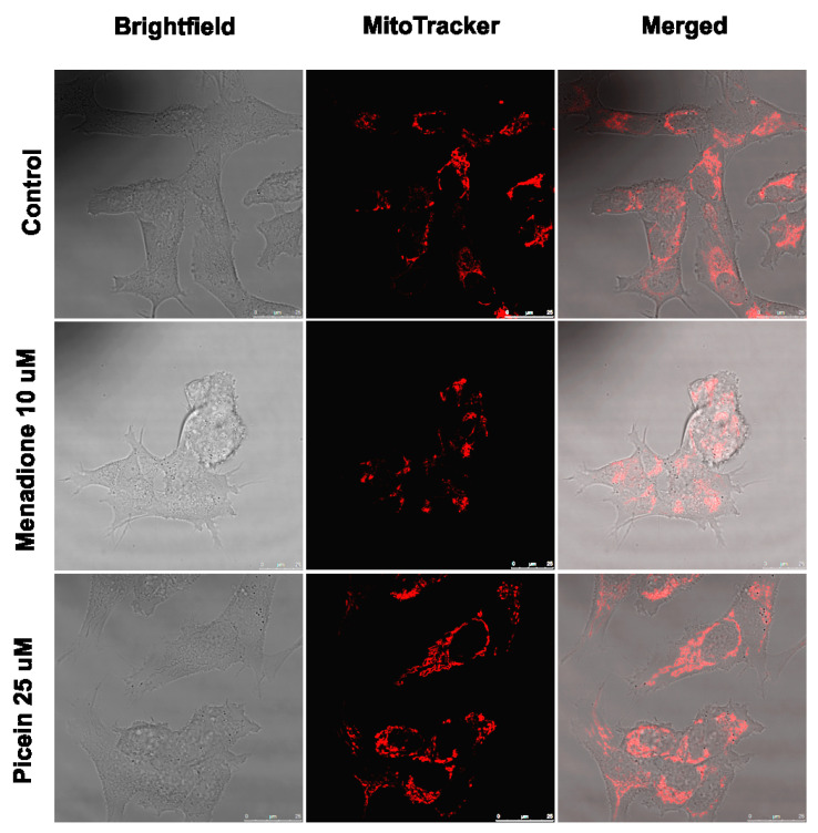
Figure 7.
Live cell microscopy of human SH-SY5Y neuroblastoma cells immediately after menadione (MQ) exposure and follow-up treatment with picein. All images were taken with a 63X water objective. Images were taken in brightfield and Mitotraker mode. Further merged these images to see the morphological differences among control, menadione (10 µM) and follow-up treatment with picein (25 µM).

