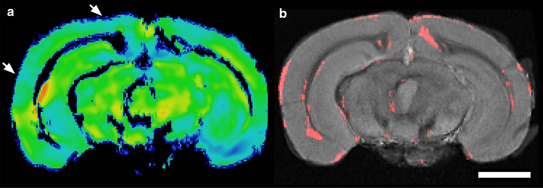
Fig. 3.

Voxel-based morphometry (VBM) and regional brain analysis. VBM was performed using high resolution ex vivo T2*W MRI, demonstrating a statistical reduction in grey matter volume, particularly in the cerebral cortex. a The combined grey matter mask and Jacobian deformation field for an individual old mouse used for VBM analysis after linear and non-linear co-registration and spatial normalization in ANTs. Intensity is mapped to deformation, where red and yellow (positive) or green and blue (negative), respectively, progressively indicated morphologic changes in a representative old-aged mouse. White arrows are representative areas of cortical thinning. b A grey-scale template is shown, created from multiple co-registered, normalized, and intensity averaged young mouse brains. The areas where the voxels are significantly different in the young from the old mouse brains, as a combination of the Jacobian Field and grey-matter mask, are overlaid in red, with p < 0.05 (t-test). The white bar represents a scale bar with size of 2 mm
