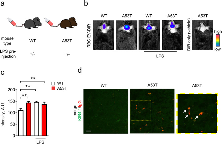
Fig. 1.
RBC-EVs can cross BBB in A53T mice. a Schematic representation showing the injection strategy. Briefly, RBC-EVs derived from control subjects were injected into WT or A53T mice in the presence or absence of LPS pre-administration. b-c Representative images and quantification of fluorescence signal of DiR-labeled RBC-EVs measured 3 h after injection. (n = 5 for WT injected with DiR-labeled RBC-EVs, n = 3 for other groups; means + S.E.M; **p < 0.001 by ordinary Two-way ANOVA test and Sidak’s multiple comparisons post-test). d Representative images of mouse brain slice from WT or A53T mouse (striatum) labeled with antibodies against mouse serum IgG and Kir4.1. Note that IgG signal can be only detected in A53T mice, and is co-localized with Kir4.1 labeled area

