Fig. 6.
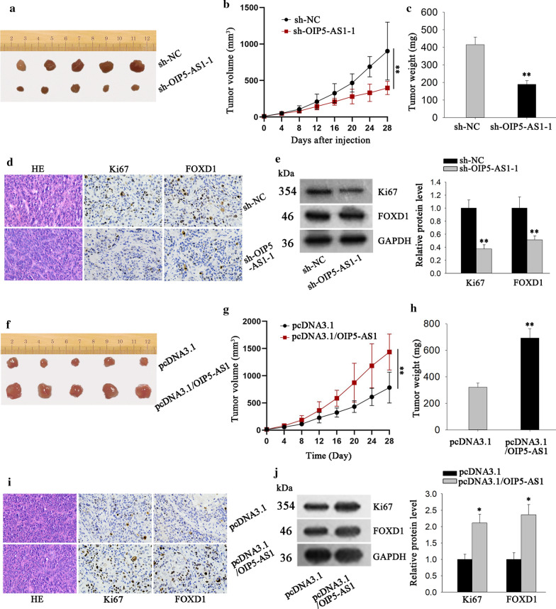
OIP5-ASI facilitates tumor growth in vivo. a The representative images of tumors from mice injected with OIP5-AS1-silenced PANC-1 cells or the control cells was shown. b The volume of above tumors was detected (**P < 0.01). c The weight of above tumors was quantified. d Immunohistochemistry was operated to measure Ki67 and FOXD1 expression in tumors from two groups. e Western blot assay was adopted to examine the protein level of Ki67 and FOXD1 (**P < 0.01). f–h Representative images, volume and weight of tumors derived from mice after inoculating CFPAC-1 cells with or without OIP5-AS1 overexpression (**P < 0.01). i, j The expression of Ki67 and FOXD1 in tumors in Fig. 6f was evaluated by immunohistochemistry assay and western blot (*P < 0.05). *P < 0.05, **P < 0.01 was considered statistically significant

