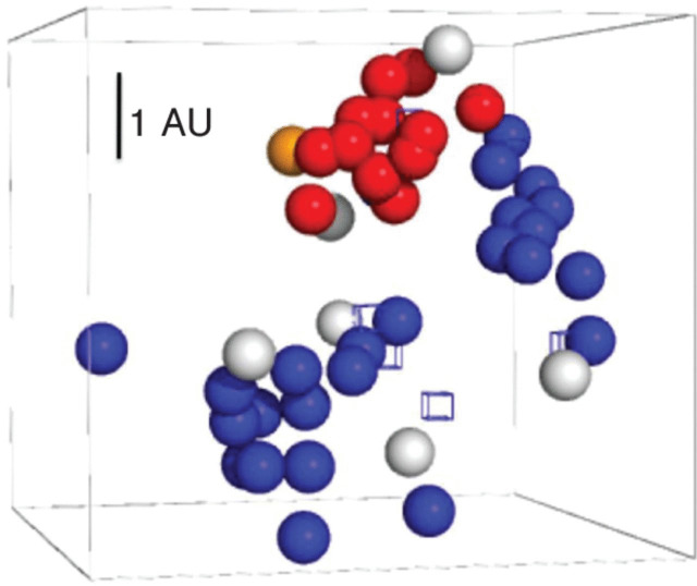
Fig. 4.
Three-dimensional antigenic map of FMDV serotype A isolates (see Fig. 2). Viruses are coloured by amino acid at position 149: proline (red), A/IRQ/24/64 (orange), others (blue), not sequenced due to confidentiality agreement (white). The cubes indicate the different sera.

