Figure 1.
TRS33 Is Required for Normal Subcellular Localization of TRS120-GFP.
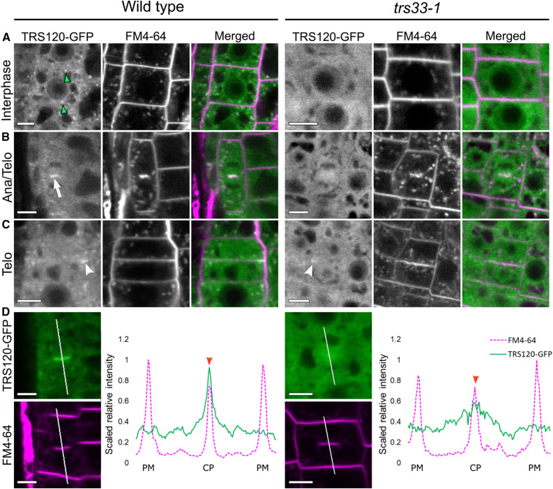
Live imaging of TRS120-GFP (green) and FM4-64 (magenta) in roots of TRS120:TRS120-GFP plants.
(A) Cells at interphase show TRS120-GFP enriched at endomembrane compartments (green arrowheads) in the wild type, but not in trs33-1, where only a cytosolic haze can be seen.
(B) During early stages of cytokinesis (cell plate initiation and biogenesis), TRS120-GFP is enriched at the cell plate (white arrow) in the wild type, but not in trs33-1. Ana, anaphase; Telo, telophase.
(C) During late stages of cytokinesis (cell plate insertion and maturation), TRS120-GFP reorganizes to the leading edges of the cell plate (white arrowhead) in the wild type. By contrast, only a weak and relatively diffuse TRS120-GFP signal can be detected at the leading edges of the cell plate in the trs33-1 mutant (white arrowhead). Telo, telophase.
(D) Line graphs depicting scaled relative fluorescence intensities. A sharp peak is seen at the cell plate (CP) in the wild type (red arrowhead), but not in trs33-1. PM, plasma membrane.
At least 10 seedlings were imaged per marker line. n = 8 for wild type, n = 7 trs33-1 for cytokinetic cells. Bars = 5 μm.

