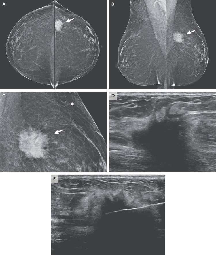
Figure 1. Imaging Studies of the Breast.
Bilateral mammograms obtained from the craniocaudal and mediolateral oblique views (Panels A and B, respectively) show a mass in the left breast underlying the skin marker (arrows). At higher magnification (Panel C), the mass appears irregular and spiculated (arrow). An ultrasound image (Panel D) shows a solid, irregular mass, measuring 3.1 cm by 1.5 cm by 1.2 cm. An image obtained during core-needle biopsy under ultrasonographic guidance (Panel E) shows the needle positioned within the mass.

