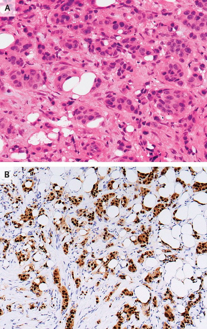
Figure 2. Core-Needle Biopsy Specimens of the Breast.
Hematoxylin and eosin staining of a tissue core (Panel A) shows invasive ductal carcinoma. Immunohistochemical staining (Panel B) shows invasive carcinoma cells that are strongly and diffusely positive for estrogen receptor and progesterone receptor.

