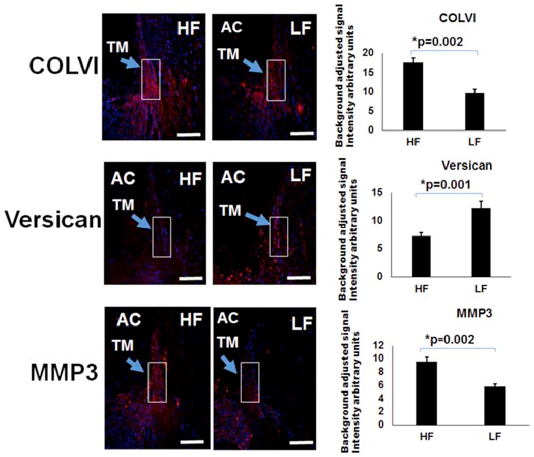
Figure 3. Differences in COLVI, Versican, and MMP3 in Segmental TM Outflow Regions by Immunofluorescence.

Immunofluorescence was performed for COLVI, versican, and MMP3. COLVI antibodies were unsuitable for Western blot. Using bead-based segmental outflow assessment methods, prior research had demonstrated elevated MMP3 and COLVI levels in HF regions and increased versican in LF regions (Keller et al., 2011; Vranka et al., 2015). Using aqueous angiography-determined angle sections, elevated COLVI was demonstrated in HF sections, versican in LF sections, and MMP3 in HF sections. White boxes demonstrate regions of interest that were used for quantification. Relative fluorescence was quantitatively evaluated using six sections each from at least three different donors and compared with an unpaired two-tailed Student’s t-test. Scale bar = 100 microns. HF = high-flow and LF = low-flow. COLVI = collagen 6 and MMP3 = matrix metalloproteinase 3.
