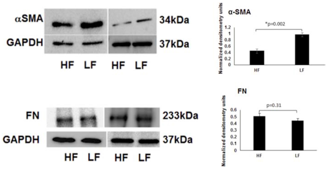
Figure 5. αSMA and Fibronectin (FN) Protein Expression in Segmental TM Outflow Regions by Western Blot.

Using aqueous angiography-determined TM tissue, elevated αSMA was demonstrated in LF regions. However, FN levels were not statistically significantly different comparing LF and HF regions. Sample size = 3 different eyes for each condition. Conditions compared by unpaired two-tailed Student’s T-test and p < 0.05 was considered statistically significant. HF = high-flow and LF = low-flow. FN = fibronectin and αSMA = alpha-smooth muscle actin.
