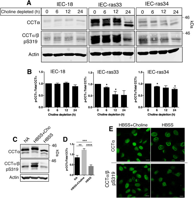
FIGURE 6:
Choline depletion induces dephosphorylation of CCTα S319 in rat intestinal epithelial cells. (A) IEC-18, IEC-ras33, and IEC-ras34 were cultured in choline-free DMEM with 5% dialyzed FBS for up to 24 h. Whole-cell lysates were immunoblotted with the primary antibodies against CCTα, CCTα/β-p-S319, and actin. (B) Phosphorylation of S319 was quantified relative to CCTα protein and expressed relative to cells that were not choline depleted (0 h). Results are the mean and SEM of three experiments and statistical comparison was with nondepleted controls (0 h). (C) IEC-ras34 cells were cultured in IEC-MEM (NA, no addition), HBSS with 50 µM choline, or HBSS for 6 h. Whole-cell lysates were resolved by SDS–PAGE and immunoblotted with CCTα, CCTα/β-p-S319, and actin antibodies. (D) Phosphorylation of S319 was normalized to CCTα protein and expressed relative to control (NA). Results are the mean and SEM of five experiments. (E) IEC-ras34 cultured in HBSS without or with choline for 6 h were immunostained with CCTα or CCTα/β-pS319 primary antibodies followed by an Alexa Fluor–488 secondary antibody. Confocal images are shown (0.8-μm sections); bar, 10 μm.

