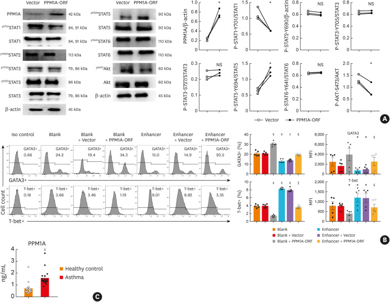
Fig. 6. PPM1A regulated Th2 differentiation through STAT and AKT signaling pathways. (A) PPM1A expression was determined by western blot analysis in cells transfected with either pcMV6-entry (vector) or PPM1A-ORF. STAT and AKT were analyzed by western blot. β-actin was included as an internal control (n = 3–6). PPM1A-ORF vs. vector; (B) Flow cytometry analysis of GATA-3 expression or T-bet expression in Th2-polarized CD4+ T cells infected with miR-1165-3p enhancer lentivirus and/or transfected with PPM1A-ORF. Data are representative of independent experiments with similar results (n = 3–6) (C) PPM1A in the plasma of the asthmatic and control groups was quantified (n = 18–20).
PPM1A, protein phosphatase, Mg2+/Mn2+-dependent 1A; STAT, signal transducer and activator of transcription; NS, not significant; miR-1165-3p, microRNA-1165-3p.
*P < 0.05, †P < 0.01, ‡P < 0.001 vs. blank group; §P < 0.05, ‖P < 0.01, enhancer + PPM1A-ORF vs. enhancer + vector.

