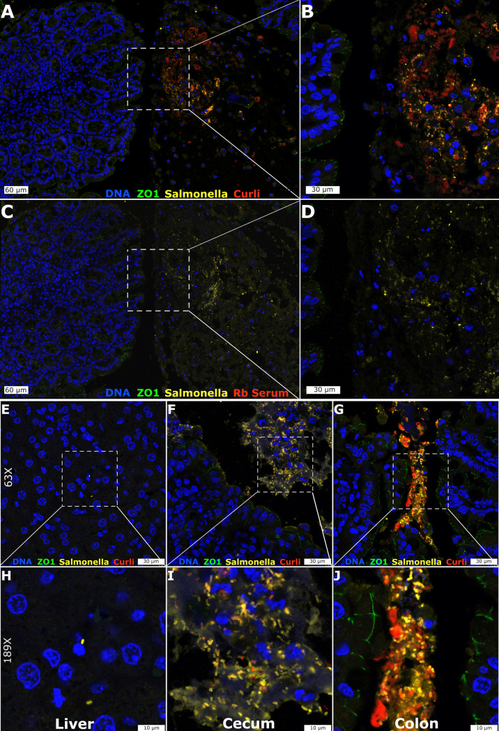
Fig 1. Immunofluorescent detection of curli produced by S. Typhimurium in the mouse gut.
Representative confocal images at 28x (A, C), 63x (E-G), 72x (B, D) and 189x (H-J) are shown for the S. Typhimurium-infected mouse Cecum (A-D, F, I), Liver (E, H), and Colon (G, J). Multicolor immunofluorescent staining was directed at ZO-1 (green), Salmonella (yellow) and Curli (red) along with DAPI counterstain (blue). Naïve rabbit serum was used as a control for non-specific staining (C, D).

