
Figure 1. Chromosome bridge breakage requires actomyosin contractility.
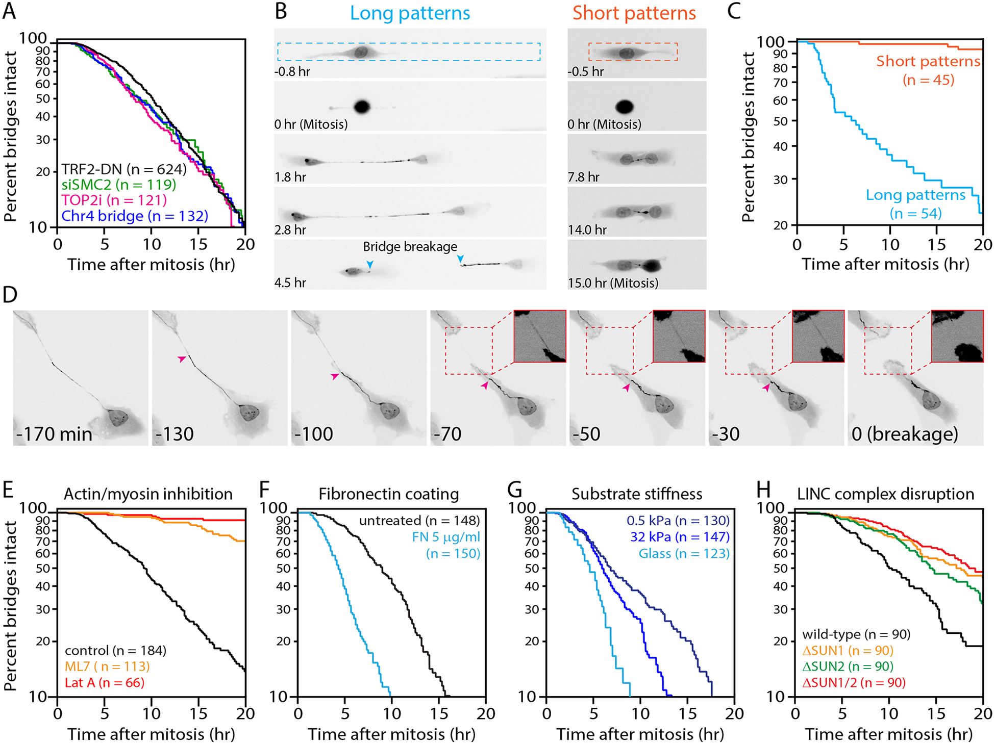
(A) Indistinguishable chromosome bridge lifetimes observed with different methods for bridge induction. Shown are bridge lifetimes (time from bridge formation until breakage or mitotic entry). Bridges were visualized with GFP-BAF and generated by: inducible TRF2-DN (n=624 bridges analyzed), condensin partial knockdown (siSMC2, n=119), low-dose ICRF-193 (n=121), or inducible CRISPR/Cas9 cutting of Chr4 subtelomere (n=132). These mean bridge lifetimes are not significantly different (p=0.14, one-way ANOVA).
(B) Extension of chromosome bridges is required for their breakage. Time-lapse images (GFP-BAF) of cells with bridges on “long” (20×300 μm) or “short” (20×100 μm) fibronectin micropatterns. Bridge length does not exceed ~50 μm on short patterns. Dashed lines: micropattern borders. Teal arrowheads: broken bridge ends. Timestamp is relative to completion of the previous mitosis.
(C) Quantification from (B): bridge lifetime on short (n=45) and long (n=54) micropatterns (p<0.0001, Mann-Whitney).
(D) Representative chromosome bridge breakage event. Prior to breakage, there is apparent non-uniform stretching of the bridge (GFP-BAF). Magenta arrowhead: a transition between “taut” and “slack” regions of the bridge. The taut region progressively stretches, the slack region progressively retracts, and breakage occurs in the taut region. Inset images: high contrast of the taut region (dashed red boxes) before and after breakage. Timestamp is relative to bridge breakage.
(E) Actomyosin contractility is required for bridge breakage. Cells were allowed to divide and form bridges before exchange into drug medium (see Fig. S5D for scheme). Plot shows bridge lifetimes with actin disruption (LatA, n=66), myosin-II inhibition (ML7, n=113), and control (DMSO, n=184).
(F) Increased cellular contractility decreases bridge lifetime. Bridge lifetimes on untreated glass (n=148) or fibronectin (FN)-coated glass (n=150).
(G) Bridge breakage timing depends on substrate stiffness: glass (>106 kPa, n=123), stiff gel (32 kPa, n=147), and soft gel (0.5 kPa, n=130). All substrates were coated with 5 μg/ml fibronectin.
(H) Partial requirement of LINC complex for bridge breakage: wild-type (n=90), ΔSUN1 (n=90), ΔSUN2 (n=90), and ΔSUN1/ΔSUN2 (n=90) RPE-1 cells.