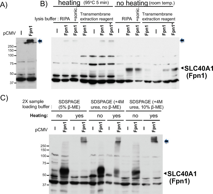
Fig 2. Comparison in Fpn1 (SLC40A1) western blotting between heated and unheated protein samples from Fpn1 transfected cells.
A) WCL in RIPA buffer was prepared from HEK293 cells transfected with pCMV or pCVMFpn1. 25ug of the WCL mixed with 2X SDSPAGE sample loading buffer was heated at 95°C for 5min and subjected to Fpn1 western blotting. The arrow indicates the Fpn1 protein stuck on the top of the separation gel (also in B and C). B) WCL in RIPA buffer, in RIPA plus sonication for 10 seconds three times in ice-water, or in transmembrane protein extraction reagent (Five Photon Biochemicals) was prepared from HEK293 cells transfected with pCMV or pCVMFpn1(6ug, *Fpn1: 12ug DNA). 15ug of the WCLs mixed with 2X SDSPAGE sample loading buffer were heated at 95°C for 5min or not heated (at room temperature), and subjected to Fpn1 western blotting. The arrowhead indicates the transfected Fpn1 band. C) WCLs in RIPA buffer from HEK293 cells transfected with pCMV or pCVMFpn1 were mixed with 2X SDSPAGE sample loading buffer containing 5% β-mercaptoethanol (regular), 4M urea, or both 5% β-mercaptoethanol and 4M urea. One sample set was heated at 95°C 5min, another set was not heated (at room temperature), and subjected to Fpn1 western blotting. Positions of molecular weight protein size marker are indicated on the left. The experiments were repeated three times and the representative western blots are shown.

