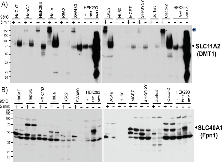
Fig 4. Comparison in DMT1 and Fpn1 western blotting between heated and unheated protein samples from 12 human cell lines.
25ug of WCLs in RIPA buffer from indicated 12 human cell lines were mixed with 2X SDSPAGE sample loading buffer, either heated at 95°C for 5min or not heated (at room temperature), and subjected to A) DMT1 and B) Fpn1 western blotting. As a control, 10ug of non-heated WCL from HEK293 cells transfected with pCMV, pCMVDMT1, or pCMVFpn1 was loaded. The arrowheads indicate the positions of transfected DMT1 and Fpn1 bands. In A), the arrow indicates the DMT1 protein stuck on the top of the separation gel. The experiments were repeated four times and the representative western blots are shown.

