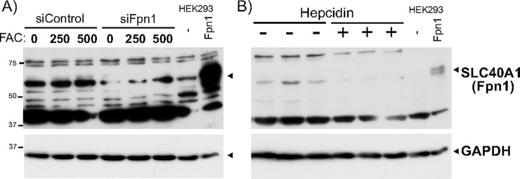
Fig 5. Verification of the band specific to endogenous Fpn1 protein.
A) Caco-2 cells were transfected with Control- or Fpn1-siRNA using Lipofectamine RNAiMax for 18hr, followed by treatment with FAC at the final 250 and 500uM for additional 8hr in the presence of siRNA. Unheated Caco-2 WCLs in RIPA buffer along with unheated WCL from HEK293 cells transfected with pCMV or pCVMFpn1 were analyzed by western blotting for Fpn1 (top) followed by incubation with anti-GAPDH antibody. B) Caco-2 cells were untreated or treated with 500nM hepcidin (3 plates each) for 22h. 20ug of three-independent hepcidin (-) and hepcidin (+) WCLs in RIPA buffer were mixed with 2X SDSPAGE sample loading buffer, unheated, and subjected to Western blotting with the anti-Fpn1 antibody. Unheated WCL from HEK293 cells transfected with pCMV or pCVMFpn1 was loaded as a control. The membrane was incubated with the anti-GAPDH antibody to assess an equal amount of sample loading. The experiments were repeated five times and the representative western blots are shown.

