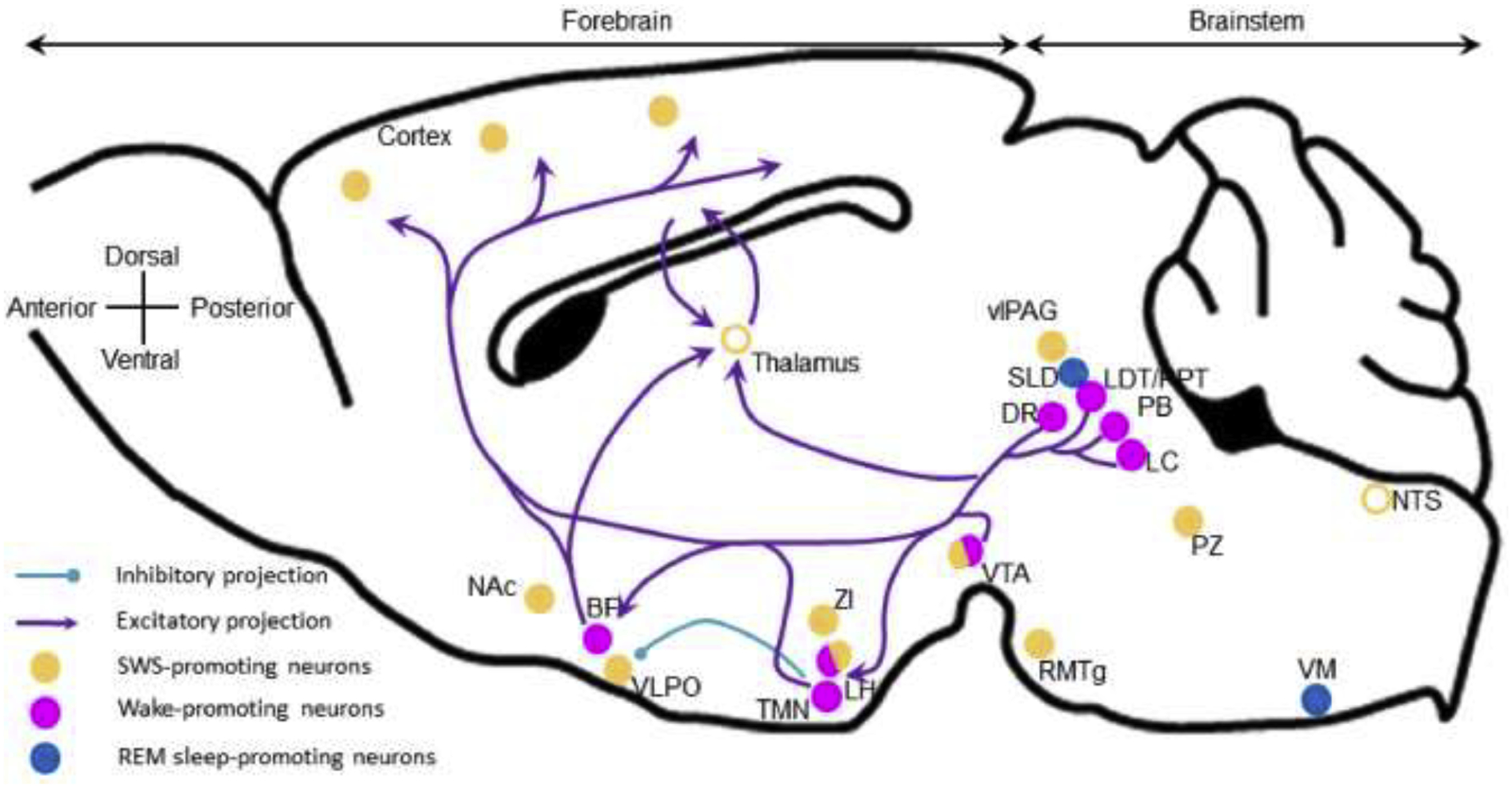
Figure 1: Wake-promoting nuclei and projections. Sagittal plane of a mouse brain.

Wake-promoting neurons are located in the brainstem and in the forebrain, and project to the cortex to actively promote cortical activation and wakefulness. Histaminergic neurons located in the TMN actively inhibit VLPO sleep-promoting neurons. Open circles, brain areas contributing to sleep control but not sleep-promoting per se. BF, basal forebrain; DR, dorsal raphe; LDT/PPT, laterodorsal and pedunculopontine tegmental nuclei; LH, lateral hypothalamus; LC, locus coeruleus; NAc, nucleus accumbens; NTS, nucleus of the solitary tract; PB, parabrachial nucleus; PZ, parafacial zone; RMTg, rostromedial tegmental nucleus; SLD, sublaterodorsal nucleus; TMN, tuberomamillary nucleus; vlPAG, ventrolateral periaqueductal gray; VLPO, ventrolateral preoptic area; VM, ventral medulla; VTA, ventral tegmental area; ZI, zona incerta.
