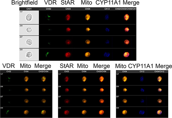
Figure 3.
Co-localization of VDR, StAR and CYP11A1 with mitochondria in human keratinocytes.
Fixed and permeabilized HaCaT cells (human keratinocytes) were stained for expression of VDR (Ch2:green), StAR (Ch3:red), mitochondria (Ch4:Orange) and CYP11A1 (Ch12:blue) and analyzed using an ImageStream II (Amms, Seattle, WA, USA) cytometer as described previously144. The composite image of four different cells (upper panel) shows that all of the StAR colocalizes with mitochondria. VDR and CYP11A1 have different subcellular distributions but both of them are partially found colocalized with mitochondria. This is observed with greater clarity when the same four cells are analyzed for VDR and mitochondria (lower left panel), StAR and mitochondria (lower middle panel) and CYP11A1 and mitochondria (lower right panel). HaCaT cells were detached and processed as previously described52. The cells were fixed, permeabilized and stained with antibodies to VDR (Santa Cruz; Dallas, TX, USA), CYP11A1 (Cell signaling technology; Danvers, MA, USA), StAR (Santa Cruz; Dallas, TX, USA), and Mitotracker Red (10 nM - CMX Ros Invitrogen; Carlsbad, CA, USA) as described previously144. Data were analyzed using IDEAS software (Amms, Seattle, WA, USA).

