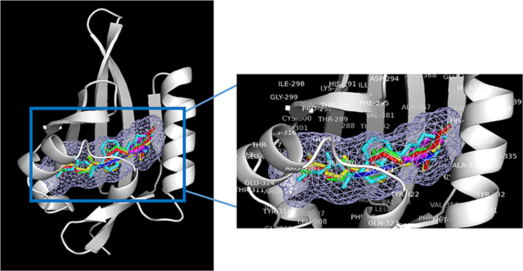
Figure 7.
Binding modes for 1α, 20S(OH)2D3(red), 1α,25(OH)2D3 (yellow), 20S(OH)D3 (green) and 20S,23R(OH)2D3 (cyan), indirubin (native ligand, blue) and indole acetic acid (native ligand, magenta) to the ligand binding domain (LBD) of AhR (in white, Homology model from previous study145). The light blue meshing area shown in the figure is the hydrophobic binding pocket in AhR. Vitamin D3 derivatives share the same ligand binding pocket with the corresponding native ligand in the LBDs for AhR.

