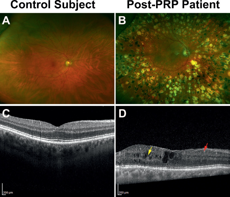
Figure 3.
Fundus and OCT images of a control participant and a diabetic patient. The diabetic retina had significant laser scars surrounding the macula (B) and showed evidence of pathologic modifications of the retinal layers (D), such as signs of disorganization of retinal inner layers (DRIL; red arrow) and cystoid changes (yellow arrow).

