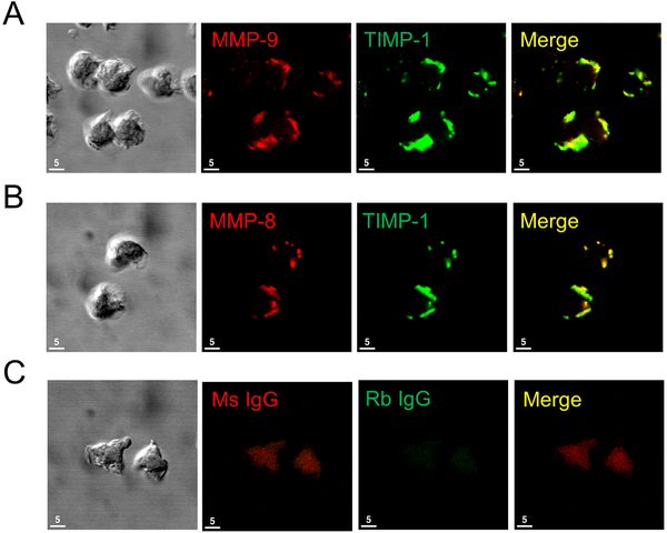
Figure 3: TIMP-1 is co-localized with MMP-8 and MMP-9 on the surface of activated human PMNs:
Human PMNs were activated at 37°C with PAF at 10−7 M for 15 min followed by fMLP at 10−7 M for 30 min. Cells were double immunostained with Alexa 546 and murine anti-MMP-9 IgG (A, second panel), or murine anti-human MMP-8 IgG (B, second panel) or non-immune murine (Ms) IgG (C, second panel) and with Alexa 488 and rabbit anti-TIMP-1 IgG (A and B third panels) or non-immune rabbit (Rb) IgG (C, third panel). The anti-MMP-8 and anti-MMP-9 IgGs used recognize both pro and active forms of these MMPs. Cells were examined using a Normarski objective (A-C, left panels) and co-localization of MMPs and TIMP-1 on the surface of the activated PMNs was assessed by confocal microscopy (see merged images in the right panels for A-C). The white bars are 5 microns in length. Note the strong co-localization of TIMP-1 with both MMP-8 and MMP-9 on the surface of activated PMNs. The results shown are representative of at least 3 different PMN preparations.

