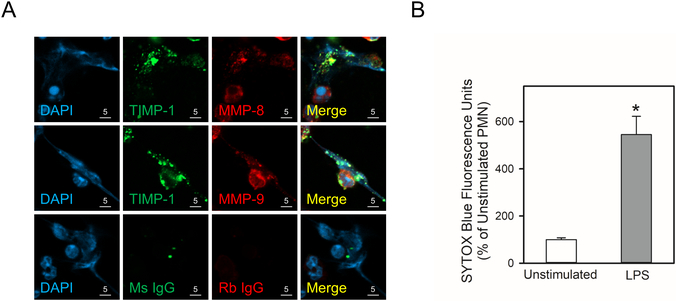
Figure 9: TIMP-1 is co-localized with MMP-8 and MMP-9 in PMN extracellular traps (NETs):
In A and B, human PMNs were incubated with 1 μg/mL LPS for 4 h at 37°C to induce NET formation, or without LPS as a control. In A, the cells were fixed and then double immunostained with a green fluorophore for TIMP-1 (second panels) and a red fluorophore for MMP-8 or MMP-9 (third panels). Nuclei were counterstained blue with 4’,6-diamidino-2-phenylindole (DAPI). Co-localization of TIMP-1 and MMPs in PMN-associated NETs was assessed by confocal microscopy (see merged images in the fourth panels). The results shown are representative of 4 different PMN preparations. In B, NET induction by LPS was quantified by staining the samples for extracellular DNA with SYTOX™ Blue Nucleic Acid Stain, and quantifying the staining as described in Methods. Data are mean + SEM; n = 4 experiments. Data were analyzed using the Mann-Whitney U tests. Asterisk indicates P < 0.05 compared with unstimulated cells.

