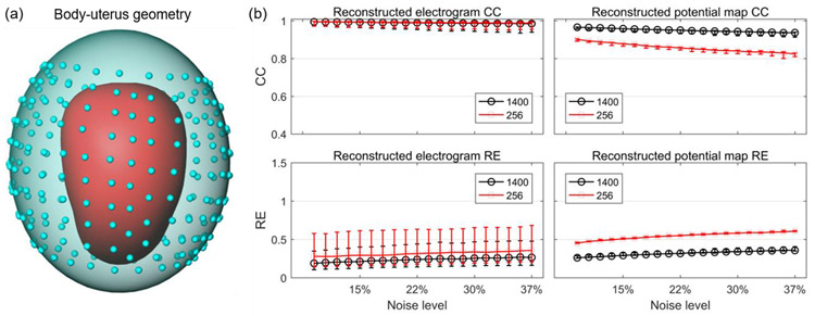
Figure 9. Reconstruction accuracy of SP method with 1400 vs. 256 body-surface sites.
a, RPI body-uterus geometry. The blue dots represent the 256 body surface sites. b, reconstruction accuracies with 1400 (black) vs. 256 (red) body surface sites. The median, first quartile, and third quartile of CC and RE values are shown by error bars.

