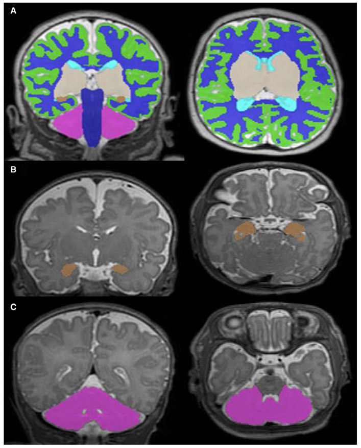
FIGURE 1.
Volumetric segmentation of T2-weighted MRI images. Coronal and axial views of the (A) total brain, (B) amygdala-hippocampus and (C) cerebellum. Legend: green = cortical grey matter, blue = white matter, grey = deep grey matter, brown = amygdala-hippocampus, pink = cerebellum, navy = brainstem and aqua = ventricles

