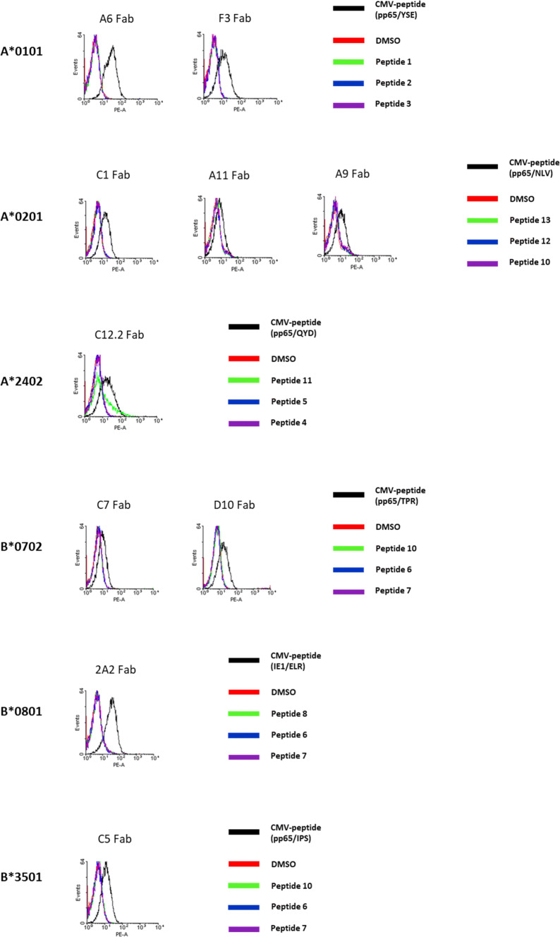
Fig. 2.
Binding assay of HLA I/HCMV-peptide-specific Fabs on LCLs. LCLs were generated by EBV infection of lymphocytes of HLA-typed donors. Peptide pulsing was done by incubation with 20 µg/ml HCMV-peptide (black line) and three control peptides (green, blue and purple lines) for 2 h at 37 °C. HCMV-peptides used for LCL pulsing are listed in Table 1 and control peptides are listed in Table S3. For staining experiments, peptide-pulsed LCL cells were incubated with 20 µg/ml biotinylated HLA I/HCMV-peptide-specific Fab antibodies for 15 min at RT. Histograms of the staining experiments are assorted from top to bottom by HLA I alleles. A*0101: HLA-restricted and HCMV-specific Fabs A6 and F3 were tested and showed specific binding to HCMV-peptide-loaded LCLs expressing the allele A*0101. Control peptides used were peptides 1, 2 and 3. A*0201: The three TCR-like Fab antibodies C1, A11 and A9 bind to HCMV-peptide-pulsed LCLs and not to the same LCLs pulsed with the control peptides 13, 12 and 10. A*2402: Binding assay of the HLA I/HCMV-specific Fab C12.2 (control peptides 11, 5 and 4) showing specific binding. B*0702: Histograms of C7 and D10 after incubation with HLA matched LCLs. Both Fabs interact only with LCLs that were pulsed with HCMV-peptide and not with same LCLs pulsed with the control peptides 10, 6 and 7. B*0801: 2A2 binds to HCMV-peptide-pulsed LCLs. As controls, the peptides 8, 6 and 7 were used. B*3501: The Fab antibody C5 shows affinity towards HCMV-peptide-pulsed LCLs (control-peptides used were 10, 6 and 7)

