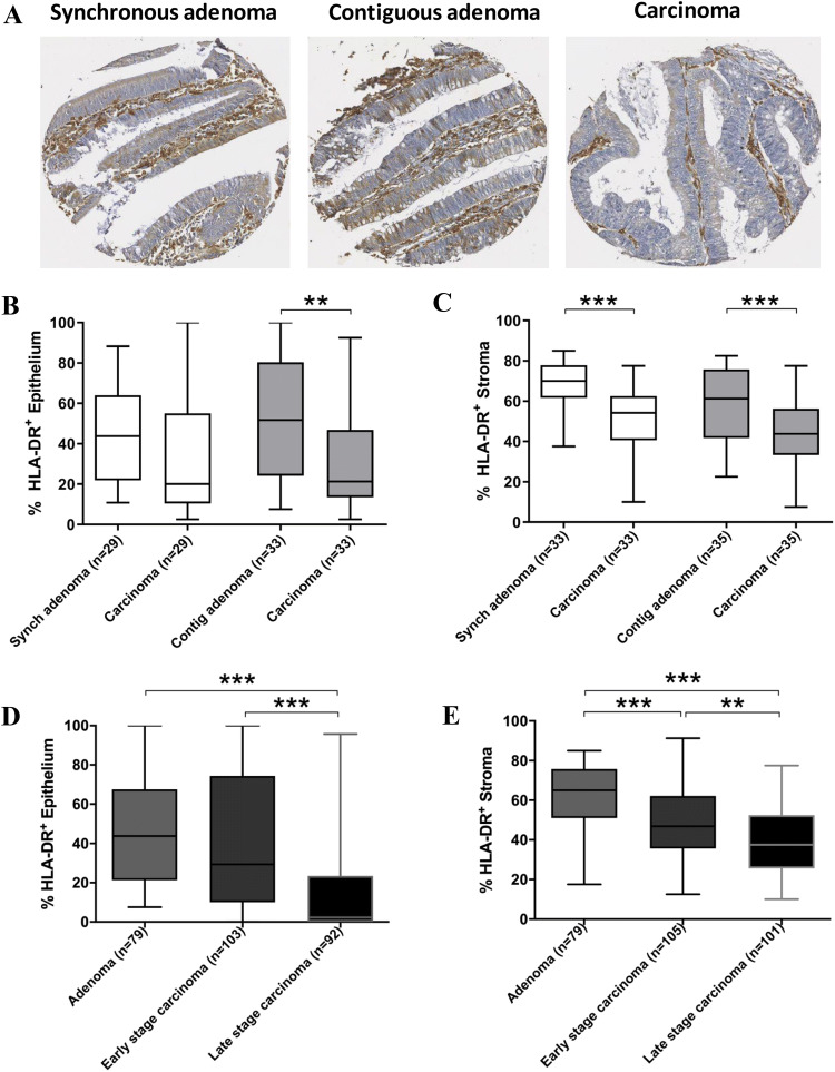
Fig. 2.
HLA-DR expression diminishes as adenomas progress to carcinomas and advance in stage. HLA-DR expression was visualised by immunohistochemistry, as shown for a single representative donor (a) and percentage positive expression was scored for colorectal tissue epithelium and stroma in a cohort of synchronous (n = 29–33) or contiguous (n = 33–35) adenomas and matched carcinomas (b, c) and adenomas (n = 79), early-stage (stage I/II, n = 103–105) and late-stage carcinomas (stage III/IV, n = 92–101) (d, e). Data were analysed using paired t test or one-way ANOVA as appropriate (Kruskal–Wallis test followed by Dunn’s multiple-comparison test). **p < 0.01, ***p < 0.001

