Figure 1.
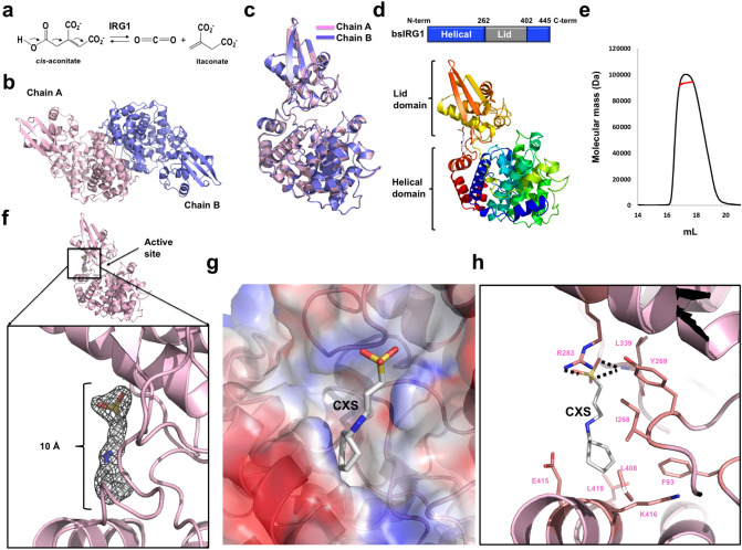
Crystal structure of Bacillus subtilis immune-responsive gene 1 (bsIRG1). (a) The production of itaconate catalyzed by IRG1 using cis-aconitate as a substrate. (b) Cartoon representation of dimeric bsIRG1. (c) Superposition of chain A and chain B of bsIRG1. (d) Domain boundary of bsIRG1. The positions of helical domain and lid domain are shown in the bar diagram shown in the upper panel. The rainbow-colored cartoon representation of monomeric bsIRG1 is shown in the lower panel. The peptide from the N- to C-termini is colored blue to red. (e) Multi-angle light scattering profile. Red line indicates the experimental molecular mass. (f) Unidentified electron density assigned as 3-cyclohexyl-1-propylsulfonic acid (CXS). 2Fo-Fc density map contoured at the 1σ level around CXS is shown. The magnified view is presented in the lower panel. (g) Electrostatic surface representation around the CXS binding pocket of bsIRG1. (h) Binding details between CXS and bsIRG1. Amino acid residues from bsIRG1, which are involved in the interaction with CXS, are labeled. Black dashed lines indicate hydrogen bonds.

