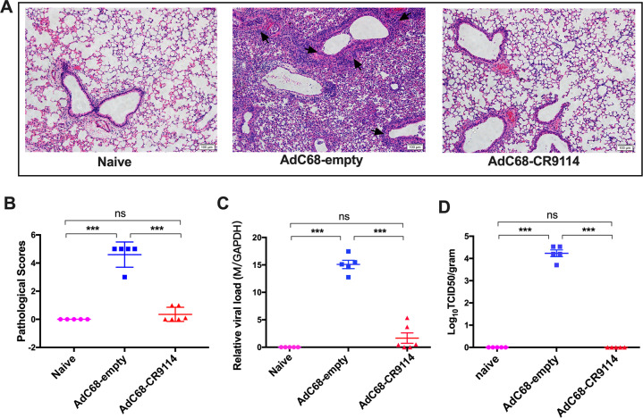
Fig. 5. Viral loads and the pathological changes in the lungs after pH1N1 challenge.
a Five days after pH1N1 challenge, mice were sacrificed, and the lung sections were stained with hematoxylin and eosin. Arrows indicated the perivascular and interstitial infiltration of inflammatory cells and the lung consolidation. b Pathological scores of the lungs 5 days after challenge. c The viral titer in the lung tissues was determined by quantitative PCR. d Lung virus titer was determined by the TCID50 assay. Five mice in each group. ***p < 0.01. ns no significance (determined using one-way ANOVA).

