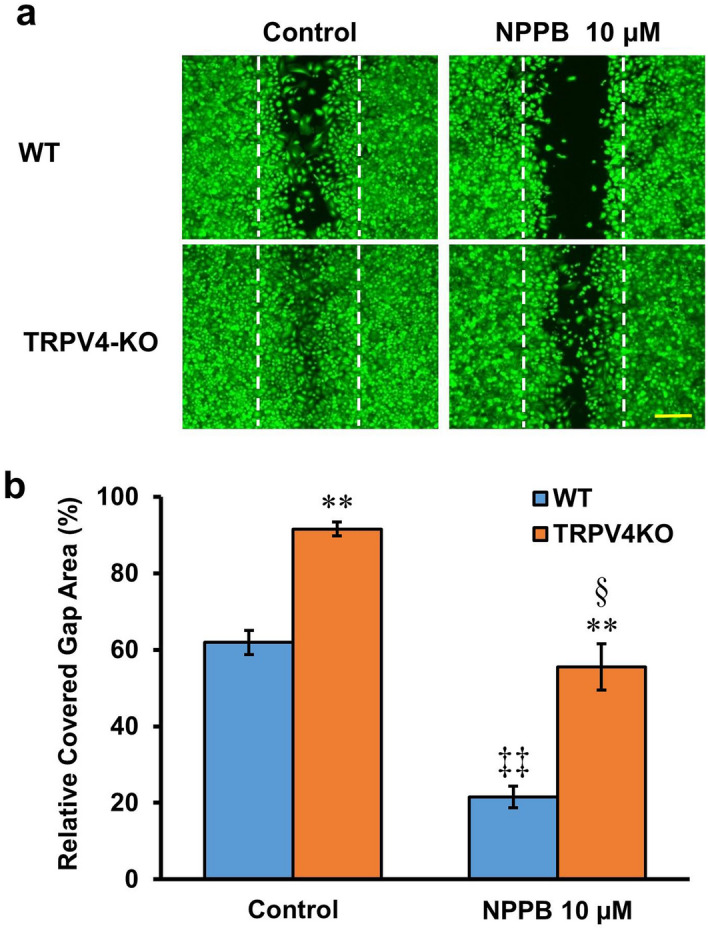
Figure 4.

Stimulation of exocytotic ATP release delays gap closure. (a) Representative images of WT and TRPV4-KO keratinocytes treated with 10 µM NPPB before staining with calcein. White dotted lines indicate the edges of the cell-free gap at 0 h after insert removal. The scale bar represents 200 μm. (b) The effect of 10 µM NPPB on percentage of covered gap area in WT and TRPV4-KO keratinocyte cultures. Data are presented as means ± SEM (n = 6–9). **p < 0.01 compared with WT, ‡‡p < 0.01 compared with WT control, §p < 0.05 compared with TRPV4-KO control; as determined by t-test.
