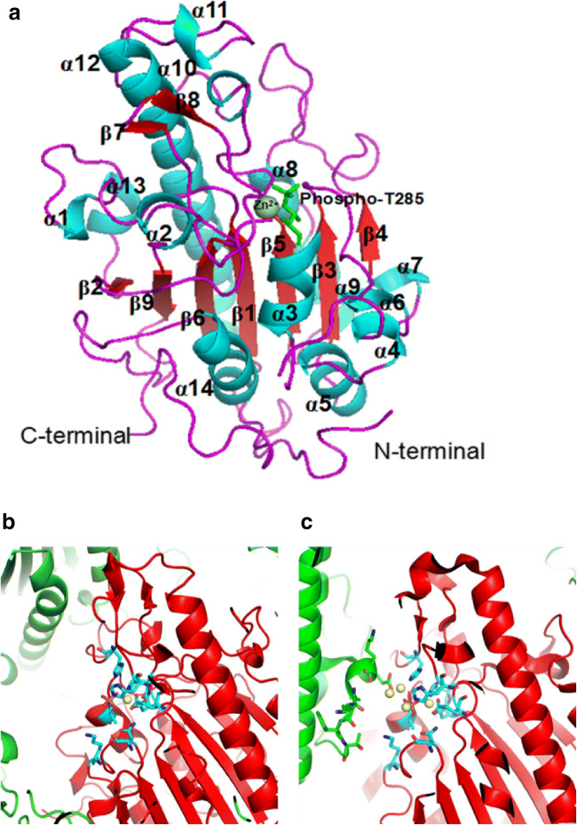
Fig. 3.
Structural characteristics of cMCR-1. a Structure of the C-terminal catalytic domain of MCR-1 (cMCR-1). The α-helices (blue), β-strands (red), loops (purple), phospho-T285 (light green), and the N- and C-termini are labeled. Zn2+ (dark green) is displayed as a sphere. b Packing diagram showing mono-zinc occupied MCR-1 in space groups P212121. c Packing diagram showing multi-zinc occupied MCR-1 in space groups P43212. The zinc ion is shown as a yellow sphere, and the catalytic core residues are presented as stick-model form (cyan). The focused MCR-1 molecule is shown in red, while the other MCR-1 molecules are shown in green

