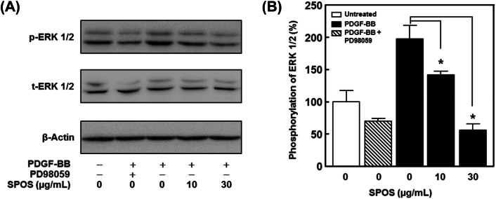
Fig. 4.
The effect of SPOS on migration and proliferation of PDGF-BB stimulated VSMCs. (A) Cell lysates were analyzed by immunoblotting with specific antibodies. (B) Statistical results obtained from panel A. The band intensities were evaluated and indicated the effect of SPOS on PDGF-BB-activated ERK1/2 phosphorylation (p-ERK). These values were normalized to total ERK1/2 (t-ERK 1/2). The basal levels of phosphorylation are deemed to be 100% (n = 3). Results are presented as mean ± standard error. Values are not significantly different based on Tukey’s multiple range test (P < 0.05)

