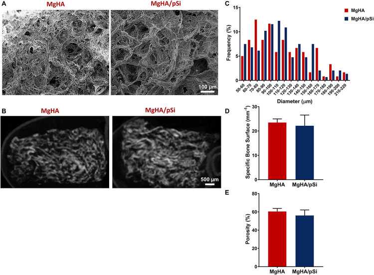
FIGURE 1.
Morphological Characterization of MgHA and MgHA/pSi scaffolds. Representative (A) SEM and (B) micro-computed tomography (μCT) images of MgHA and MgHA/pSi scaffolds. (C) Pore size distribution is shown to be similar between the two scaffolds. (D) Specific bone surface ratio and (E) percent porosity were unaffected by the addition of drug-eluting microspheres.

