Figure 5.
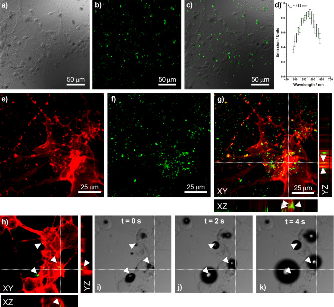
Imaging at confocal microscope of primary cultures of murine cortical neurons and astrocytes incubated with a-DG-AuNPs (700 µg/mL) for 4 h. Identification of DG-AuNPs and fluorescence: (a) DIC image (×40); (b) fluorescence image of AuNPs (λex = 488 nm); (c) merging of the images a and b showing the correspondence between the nanoparticle location and green fluorescence; (d) emission spectrum of AuNPs (λex = 488 nm). Internalization in cell of DG-AuNPs: (e) fluorescence image of astrocytes with rhodamine-stained F-actin (red); (f) fluorescence image of AuNPs (λex = 488 nm); (g) merging of images d and e, with Z-stacking lateral views showing the internalization of the AuNPs in the cell. Cell photoablation: (h) fluorescence image of neuron with rhodamine stained F-actin; (i–k) DIC images upon laser-pulsed excitation tuned at wavelength of 760 nm and power of 24 W/cm2 showing the rapid formation of bubbles in correspondence of a-DG-AuNPs.

