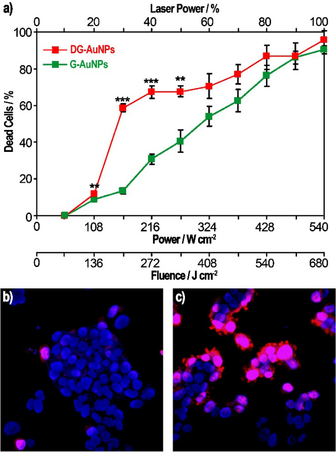
Figure 6.
Mortality of Hep G2 cells (a) after irradiation for 1.26 s at different incident powers with pulsed laser tuned at 760 nm (determined as percentage of PI-stained cells/total cells; **p < 0.01; ***p < 0.001). Representative images of Hep G2 cells, treated with Hoechst (blue) and propidium iodide (red) stains (colocalization being violet), irradiated for 1.26 s with pulsed laser tuned at wavelength of 760 nm and power of 270 W/cm2 (50% laser power) in absence of AuNPs (b) or after incubation with DG-AuNPs at concentration of 70 µg/mL for 1 h (c).

