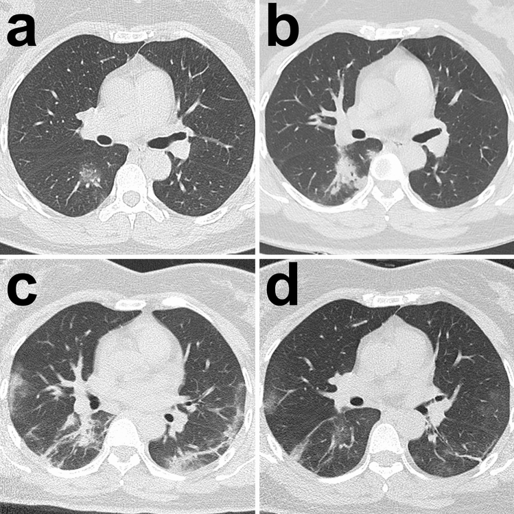
Figure 3.
Typical radiological evolution of survivors. Images from a patient presenting with sudden fever (38.8 °C) for four days. (a) At presentation (day 4), a small region of GGO was demonstrated in the right lower lobe on CT scan and the RT-PCR test was performed afterwards; (b) on admission after confirming COVID-19 (day 9), the previous GGO became more consolidated with more surrounding subpleural lesions; (c) day 13, more bilateral subpleural GGO and consolidation were observed, and the previous consolidation was partially absorbed; (d) day 18, most of the lesions were absorbed while only some residual GGO and parenchymal bands could be observed. All images have the same window level of − 600 and window width of 1,600.

