
FIGURE 4.
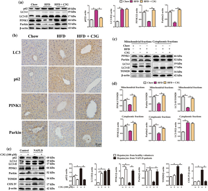
Cyanidin‐3‐O‐glucoside (C3G) restores ) high fat diet (HFD)‐impaired hepatic mitophagy. (a–d) Mice were treated as described in Figure 1 (n = 9 mice per group). (a) Protein abundance of p62, LC3, PINK1 and Parkin in the livers from different groups. (b) Representative images of hepatic IHC staining for LC3, p62, PINK1 and Parkin in different groups (original magnification 40×). (c, d) Protein levels of PINK1, Parkin and LC3 in mitochondrial or cytoplasmic fractions of the livers from different groups. (e) Protein abundance of p62, LC3, PINK1, Parkin, TOM20 and COX IV in hepatocytes from healthy controls and patients with non‐alcoholic fatty liver disease (NAFLD). Hepatocytes were treated with or without 100‐μM C3G for 12 h (n = 6). Data were expressed as the mean ± SEM. * P < 0.05