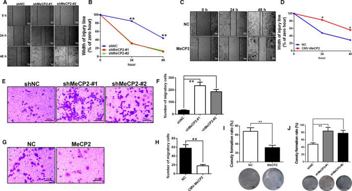
Figure 3.

MeCP2 inhibits proliferation, motility and migration in breast cancer cells. A and B, After 48 h of cells culture, the wound healing assay showed that knockdown of MeCP2 in MCF‐7 cells resulted in increased cellular motility. Scale bars = 50 µm, Magnification, 40×. C and D, Overexpression of MeCP2 in MDA‐MB‐231 cells resulted in decreased cellular motility. Scale bars = 50 µm, Magnification, 40×. E and F, Transwell assays in monolayer cultures were performed to assess tumorigenesis of MCF‐7 cells treated with shMeCP2 knockdown vectors. Scale bars = 200 µm, Magnification, 200×. G and H, MDA‐MB‐231 cells transfected with an MeCP2 overexpression vector. Scale bars = 200 µm, Magnification, 200×. I, In the colony formation assay, representative micrographs and quantitative data show the colony formation rate in MDA‐MB‐231 cells that overexpress MeCP2. Magnification, 100×. J, MCF‐7 cells that knocked down for expression of MeCP2. Magnification, 100×. All experiments were performed at least three times, and data were statistically analysed using a Student's t test (*P < .05, **P < .01 and ***P < .001 vs control)
