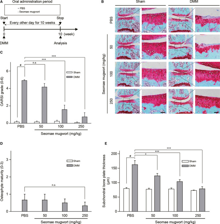
FIGURE 2.

Seomae mugwort extract suppressed the DMM‐induced cartilage degradation. A, Experimental scheme for the analysis of a DMM‐induced OA model. After DMM surgery, the mice were orally administrated the Seomae mugwort extract or PBS every other day for 10 wk. B, Ten‐week‐old C57BL/6 mice were subjected to DMM. Mice were orally administered PBS or the Seomae mugwort extract for 10 weeks after DMM. Cartilage degradation was shown by safranin O staining. Scale bar = 100 μm. C‐E, The severity of OA was evaluated by OARSI scores (C), osteophyte maturity (D) and subchondral bone plate thickness (E) at 10 weeks after DMM. Mice orally administered the Seomae mugwort extract were compared with those administered PBS. Values are the mean ± SEM as analysed by one‐way ANOVA with Bonferroni's test (n = 10 mice per group). Significant differences from the DMM group value: *P < .05, **P < .01, ***P < .001, #P < .05 compared to the control group
