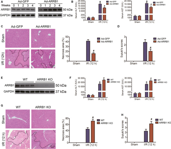
FIGURE 2.

ARRB1 protects against hepatic I/R injury. A, After virus injection, primary hepatocytes isolated at the indicated time‐points were subjected to Western blot to detect the transfection efficiency. B, Serum AST and ALT activities in Ad‐GFP or Ad‐ARRB1 mice subjected to sham or hepatic I/R for 12 h of reperfusion as indicated (n = 5‐6 per group). C, Representative images of histological H&E staining showing necrotic areas in livers from Ad‐GFP or Ad‐ARRB1 mice in the sham and I/R group at 12 h following reperfusion (magnification, ×200, n = 5‐6 per group). D, The Suzuki's scores of liver sections in C. E, Western blot analysis of ARRB1 in primary hepatocytes from WT and ARRB1 KO mice (n = 4 per group). F, The activities of AST and ALT in the serum of WT and ARRB1 KO mice challenged with sham or hepatic I/R at 12 h of reperfusion (n = 5‐6 per group). G, Representative H&E staining images, statistics showing necrotic areas of liver lobes from WT and ARRB1 KO mice in the sham and I/R group at 12 h following reperfusion (magnification, ×200, n = 5‐6 per group). H, The Suzuki's scores of liver sections in G. For B‐D, *P < .05 compared with sham group; # P < .05 compared with corresponding Ad‐GFP group. For F‐H, *P < .05 compared with sham group; # P < .05 compared with corresponding WT group. All data are presented as the mean ± SD; For C, D and G, H, significance was determined by Student's two‐tailed t test; For B and F, significance was determined by one‐way ANOVA
