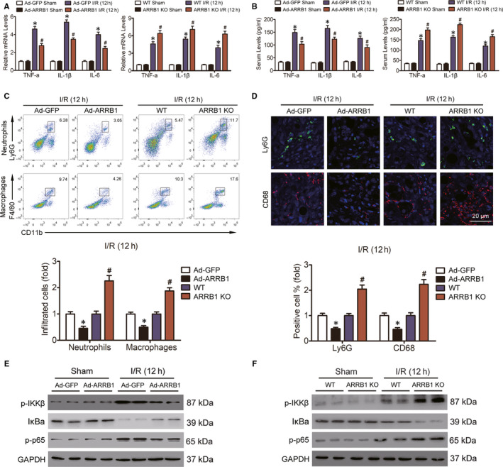
FIGURE 4.

ARRB1 restrains inflammatory response in livers during hepatic I/R injury. A, The mRNA levels of pro‐inflammatory cytokines in mouse liver and the production of them in the serum (B) of mice were measured 12 h post‐reperfusion (n = 5‐6 per group). C, Flow cytometry analysis was conducted to examine the accumulation of neutrophils and macrophages in livers from mice subjected to 12 h post‐reperfusion (n = 5‐6 per group). D, Representative immunofluorescence staining of Ly6G‐positive cells (green) and CD68‐positive cells (red) in the livers of Ad‐GFP and Ad‐ARRB1, WT and ARRB1 KO mice 12 h after I/R injury (magnification, ×400, n = 5‐6 per group). E and F, Immunoblotting analysis of the activation of NF‐κB signalling in livers from these mice 12 h post‐reperfusion. GAPDH served as the loading control. For A and B, *P < .05 compared with the sham group, # P < .05 compared with the corresponding Ad‐GFP or WT groups; for C and D, *P < .05 indicates Ad‐ARRB1 compared with Ad‐GFP, # P < .05 indicates ARRB1 KO compared with WT. All data are presented as the mean ± SD; significance was determined by one‐way ANOVA
