
FIGURE 3.
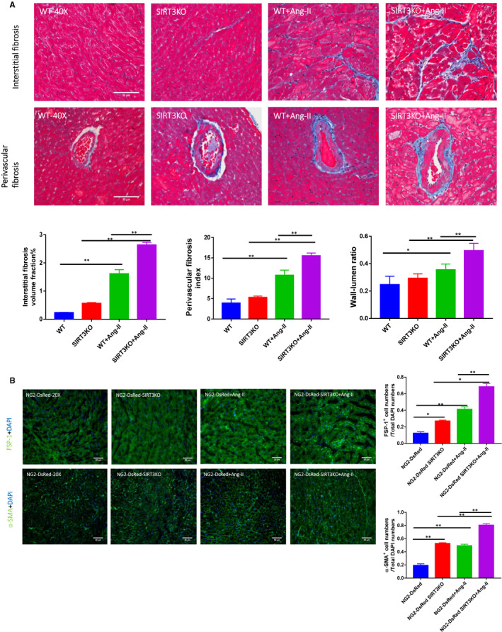
Knockout of SIRT3 accentuated Ang‐II‐induced fibrosis. A, Masson's trichrome staining showed that interstitial fibrosis was increased significantly in WT mice + Ang‐II (n = 4 mice) and knockout of SIRT3 (n = 4 mice) further enhanced this increase as compared to WT mice + Ang‐II. Knockout of SIRT3 promoted Ang‐II‐induced coronary artery remodelling. Perivascular fibrosis and increased wall‐lumen ratio were found in WT mice + Ang‐II (n = 4 mice); these alterations were accentuated by knockout of SIRT3. Mean ± SEM, *P < .05, **P < .01. B, Immunostaining revealed a significant increases in FSP‐1 and α‐SMA levels in NG2‐DsRed mice + Ang‐II (n = 4 mice), and levels of FSP‐1 and α‐SMA were further increased by knockout of SIRT3. Mean ± SEM, *P < .05, **P < .01