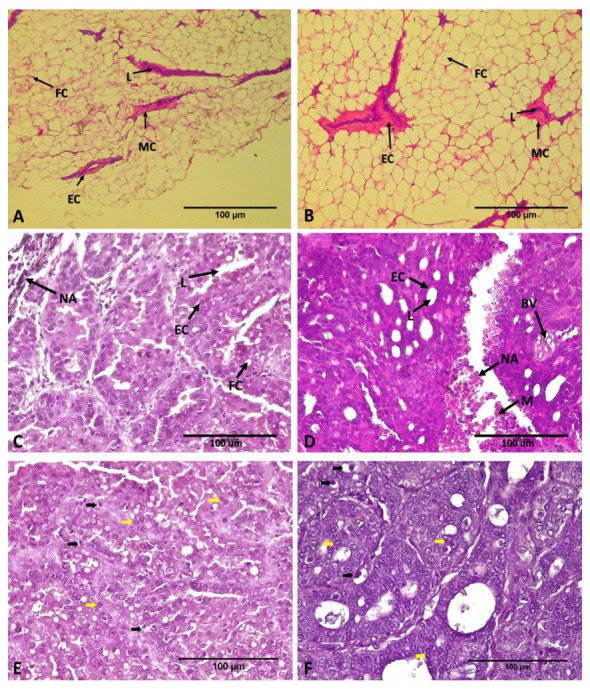
Figure 2. Histological sections of the mammary gland and DMBA-induced rat breast adenocarcinoma after treatment with ECCT exposure.

( A and B) There is no morphological change both on mammary gland tissue of none DMBA-induced rat non-EF therapy (NINT; A), and with EF therapy (NIT; B). Hematoxylin and eosin staining, magnification 100x. ( C and D) In contrast, adenocarcinoma breast tissue of DMBA induced rat non-EF therapy (INT; C) shows more massive tumor cells and fewer lumens than the tumor section with EF therapy (IT; D). Hematoxylin and eosin staining, magnification 400x. ( E and F) Mitotic figure (black arrow) and apoptotic figure (yellow arrow) on breast tumor tissues of INT and IT group, respectively. FC= Fat Cells, L=Lumens, EC=Epithelial Cells, M= Myoepithelial Cell, NA= Necrotic Area, BV= Blood Vessels.
