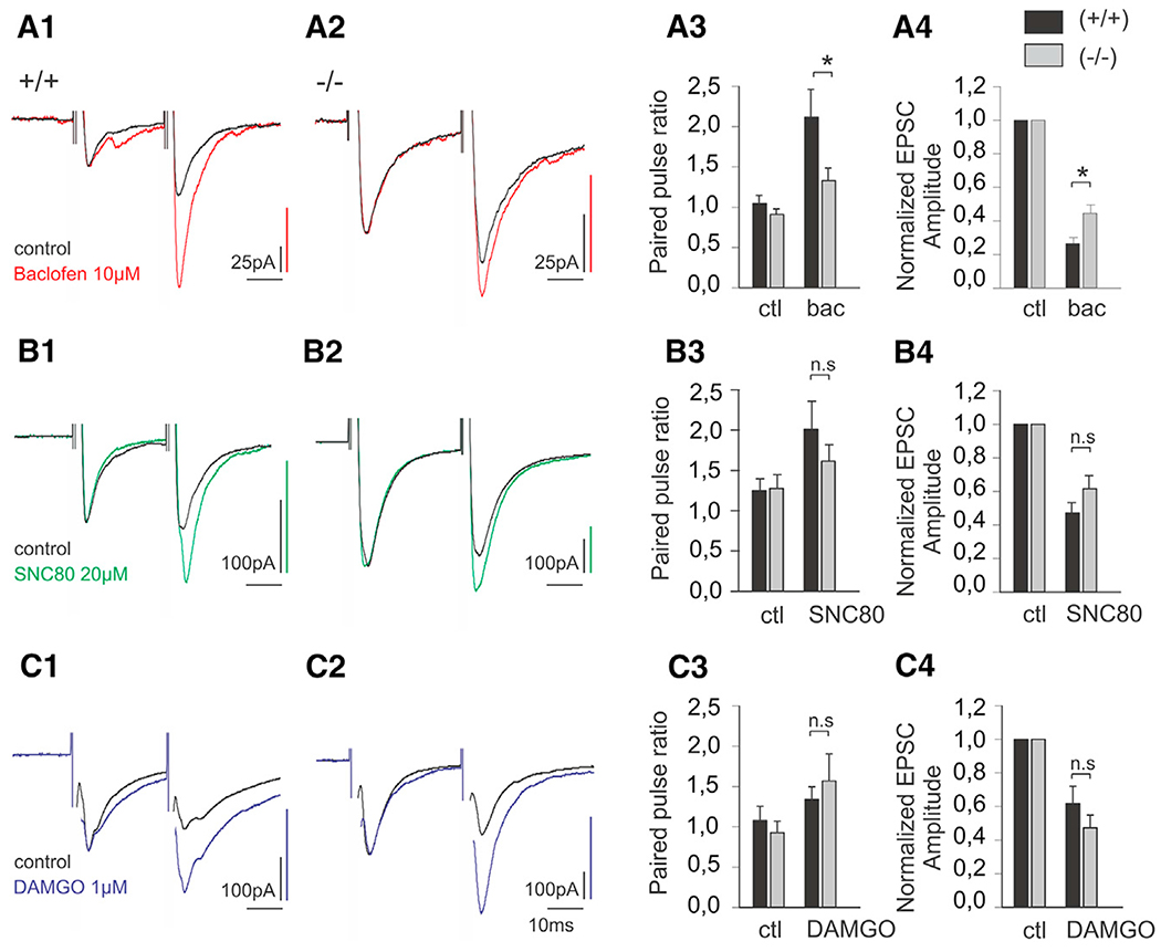
Figure 7. Modulation of Synaptic Transmission in GINIP Null Mice.
(A) Normalized whole-cell voltage clamp recordings of EPSCs evoked by a pair of electric shock on attached dorsal root in WT (A1) and GINIP−/− (A2) animal lamina II neurons, in control and following superfusion of baclofen (10 μM). (A3) Paired pulse ratio of evoked EPSCs in control and following baclofen bath application in WT and GINIP−/− mice. (A4) Normalized evoked EPSC amplitude in control and following baclofen superfusion.
(B) Normalized whole-cell voltage clamp recordings of EPSCs evoked by a pair of electric shock on attached dorsal root in WT (B1) and GINIP−/− (B2) animal lamina II neurons, in control and following superfusion of SNC80 (20 μM). (B3) Paired pulse ratio of evoked EPSCs in control and following SNC80 bath application in WT and GINIP−/− mice. (B4) Normalized evoked EPSC amplitude in control and following SNC80 superfusion.
(C) Normalized whole-cell voltage clamprecordings of EPSCs evoked by a pair of electric shock on attached dorsal root in WT (C1) and GINIP−/− (C2) animal lamina II neurons, in control and following superfusion of DAMGO (1 μM). (C3) Paired pulse ratio of evoked EPSCs in control and following DAMGO bath application in WT and GINIP−/− mice. (C4) Normalized evoked EPSC amplitude in control and following DAMGO superfusion.
Traces in A1, A2, B1, B2, C1, and C2 have been scaled in order to facilitate the comparison of PPR (*p < 0.05).

