Figure 4.
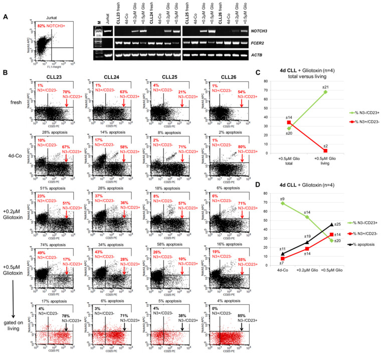
NOTCH3 and CD23 surface expression are mutually exclusive on CLL cells. (A) RT-PCR and (B) FACS showing NOTCH3 and FCER2 (CD23) expression in freshly isolated CLL cells and after 4 days in culture in relation to surface CD23 expression and spontaneous as well as gliotoxin-induced apoptosis. The T-ALL cell line Jurkat served as positive control for NOTCH3 mRNA and NOTCH3 surface expression. (C) Gating on the remaining living cells after gliotoxin treatment according to their forward/side scatter properties revealed that living CLL cells were enriched for NOTCH3-/CD23+ cells. (D) Summary of the FACS data, demonstrating a direct correlation of the percentage of NOTCH3+/CD23- and apoptotic CLL cells and an indirect correlation of these two parameters with the percentage of NOTCH3-/CD23+ CLL cells. Data presented as means (±SD).

