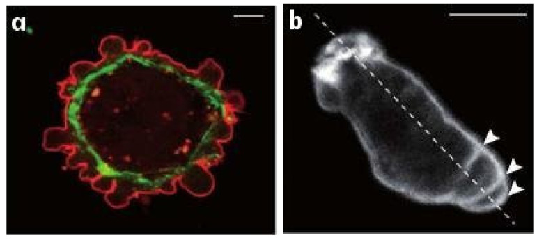
Figure 2.
(a) Blebbing on a melanoma cell: myosin (green) localizes under the blebbing membrane (red) (b) The actin cortex of a Dicty cell migrating to the lower right. Arrowheads indicate the successive blebs and arcs of the actin cortex. The scale bar is 5 m is each panel. From [26].

