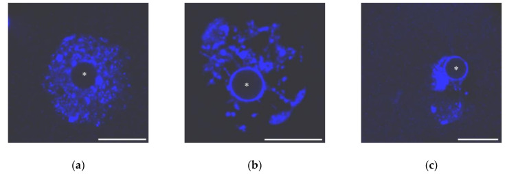
Figure 1.
The nuclei of growing mouse oocytes, demonstrating different chromatin organization as viewed after DAPI staining: (a) NSN; (b) early SN; a heterochromatin “ring” around unstained nucleolus-like body appears; and (c) late SN; chromatin is assembled in a more compact mass (karyosphere). Asterisks indicate nucleolus-like bodies. Scale bars represent 20 μm.

