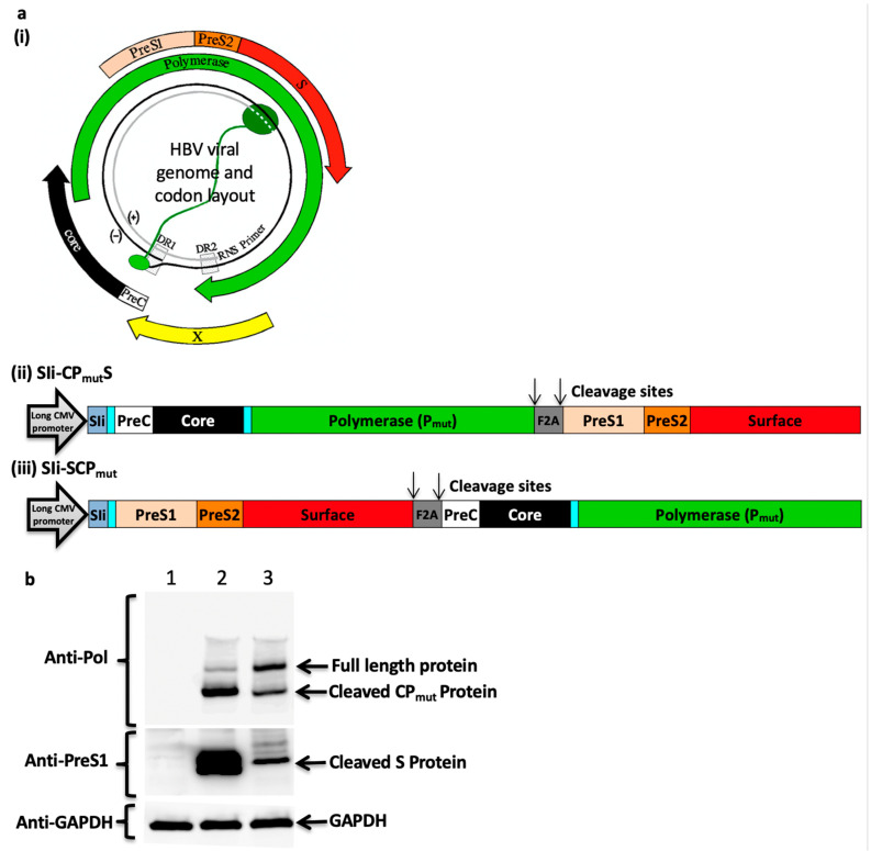
Figure 2.
HBV immunogen design and in vitro protein expression analysis. (a) HBV viral genome and codon layout (a, i) (illustration from [25]). HBV genome comprise of a partially double stranded circular DNA, of approximately 3.2 kilobase (kb) pairs. It encodes 4 coding regions; precore along with core, polymerase, surface proteins (three forms of the surface proteins, L, M, and S, where the L-form is composed on PreS1, PreS2, and S, the M-form is composed of PreS2 and S) and x-protein. HBV immunogens SIi-CPmutS (a, ii) and SIi-SCPmut (a, iii) were designed to encode HBV precore (PreC), core, polymerase (Pmut), PreS1, PreS2, and surface proteins and non-HBV regions (comprising of a truncated shark Invariant chain (SIi), two linkers and a Furin 2A (F2A) peptide sequence). The preS1/preS2/surface region was positioned at carboxy-terminus of layout 1 and at the amino-terminus of layout 2. Within the mammalian expression cassette, the immunogen sequence was placed in between a long CMV promoter and BGH poly sequence. (b) Plasmids encoding SIi-CPmutS and SIi- SCPmut were transfected into HEK293A cells. 24 h post-transfection, cells were lysed and the lysates were analysed in western blot experiments using mouse anti-HBV-PreS1 and mouse anti-HBV-Polymerase antibodies. Blots probed with mouse anti-GAPDH served as loading controls. Lane 1: cell lysate from un-transfected cells, lane 2 and lane 3: cell lysates from cells transfected with plasmids encoding SIi-CPmutS and SIi-SCPmut, respectively.

