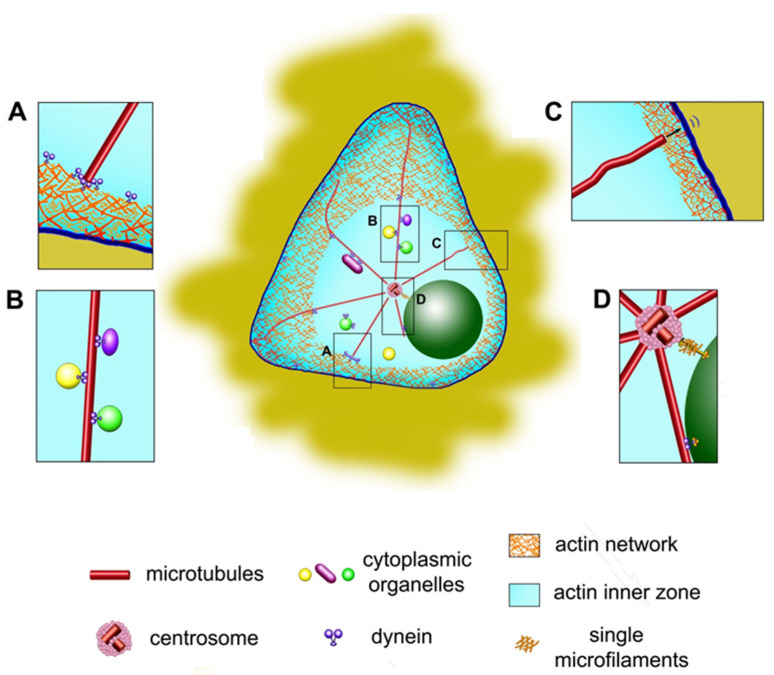
Figure 2.
Geometry and mechanism of the centrosome centering. (A)—multiple dynein molecules pulling the microtubule from the cell periphery. (B)—pulling forces applied by dynein molecules anchored at the surface of cytoplasmic organelles along the microtubule. (C)—pushing forces generated by growing microtubule plus-end. The forces applied to microtubules by the actin cortical flow are not shown on this figure (D)—links between the centrosome and the nucleus. Central panel: note that the pulling forces are applied in actin inner zone only, and curved microtubules outside it do not contribute to centering [9].

